But how does the brain know what odors the nose has detected? This is a specific version of the problem of “representation”: when we look within the brain, how is information about the outside world organized to enable sensory processing and behaviors? This is a foundational problem in neurobiology, one that has driven generations of scientists to study the brain. Although the details vary for each sensory modality, the traditional framework for understanding how the brain understands the outside world centers around the idea of the map: neurons build maps of the outside world within the brain and these maps are read by other parts of the brain to generate behaviors.

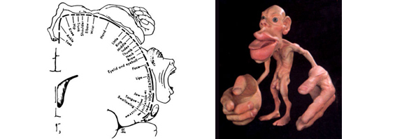
Perhaps the most famous brain map is the Penfield map – there are regions of the brain devoted to our sense of touch, and these regions are spatially organized just like the body is spatially organized, with neurons that respond to touch on the forearm residing right next to neurons that respond to touch on the elbow, and so on. This map of the body within the brain — with some imagination — looks just like a person. The important concept here is that what the brain needs to know when we are being touched is where we are being touched — and that is why the spatial relationships amongst the touch receptors in our body are maintained in the spatial relationships amongst the neurons that respond to touch in our brains. There are maps for other sensory modalities within the brain as well: the retinotopic map, in which neurons that are near each other in the visual cortex respond to spots of light that fall near each other on the retina (which in turn correspond to adjacent positions in visual space) and the tonotopic map, in which neurons that are near each other in the auditory cortex respond to similar frequencies of sound.

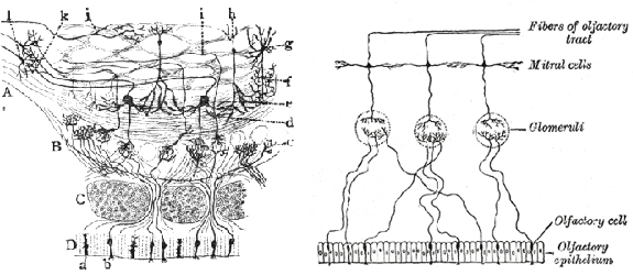
The brain also houses a map of the outside olfactory world. This map is built by the terminations of olfactory sensory neuron axons within the olfactory bulb. This map operates using three key organizational principles. First, each olfactory receptor neuron (and there are about 2 million in a mouse nose) expresses just one of the 1000 possible receptors. They make a choice to express one specific receptor early in development and then stick with that choice for their entire lifetime. Second, all of the sensory neurons that express any particular receptor send axons that converge on a single structure within each olfactory bulb called a glomerulus. Third, the spatial position within the bulb of any particular glomerulus is the same in every animal. Together, these features have an important consequence: odors activate specific sets of olfactory sensory neurons in the nose which in turn activate specific sets of glomeruli within the olfactory bulb.

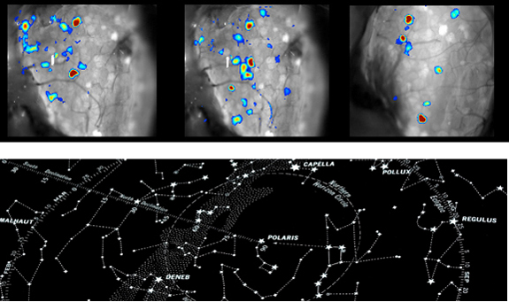
Because the specific pattern of glomeruli that any given odor activates are unique (i.e. the pattern of cherry is different from the pattern of beer), and because the spatial relationships amongst the different glomeruli are the same from animal to animal (i.e. the pattern of cherry is the same in you and me), the spatial pattern of glomeruli activated in response to an odor can be used to identify what the animal is smelling. Perhaps the best analogy is to gazing at the night sky: we can identify individual constellations (odors) by observing the conserved spatial relationships amongst the stars (glomeruli).
